The human heart is a muscular, hollow organ responsible for pumping blood throughout the body. It works continuously to supply oxygen and nutrients to tissues and to remove carbon dioxide and metabolic wastes. Structurally, the heart is well-designed to support efficient circulation.
Location and Size of the Heart
-
Located in the thoracic cavity, between the lungs
-
Slightly tilted to the left
-
About the size of a closed fist
-
Enclosed by a protective double-layered sac called the pericardium
External Structure of the Heart
The heart has a conical shape with:
-
Apex – the pointed lower end (directed downward and left)
-
Base – the broad upper part where major blood vessels are attached
Major blood vessels connected to the heart include:
-
Aorta
-
Pulmonary artery
-
Pulmonary veins
-
Superior and inferior vena cava
Internal Structure of the Heart
The heart is divided into four chambers by muscular walls (septa).
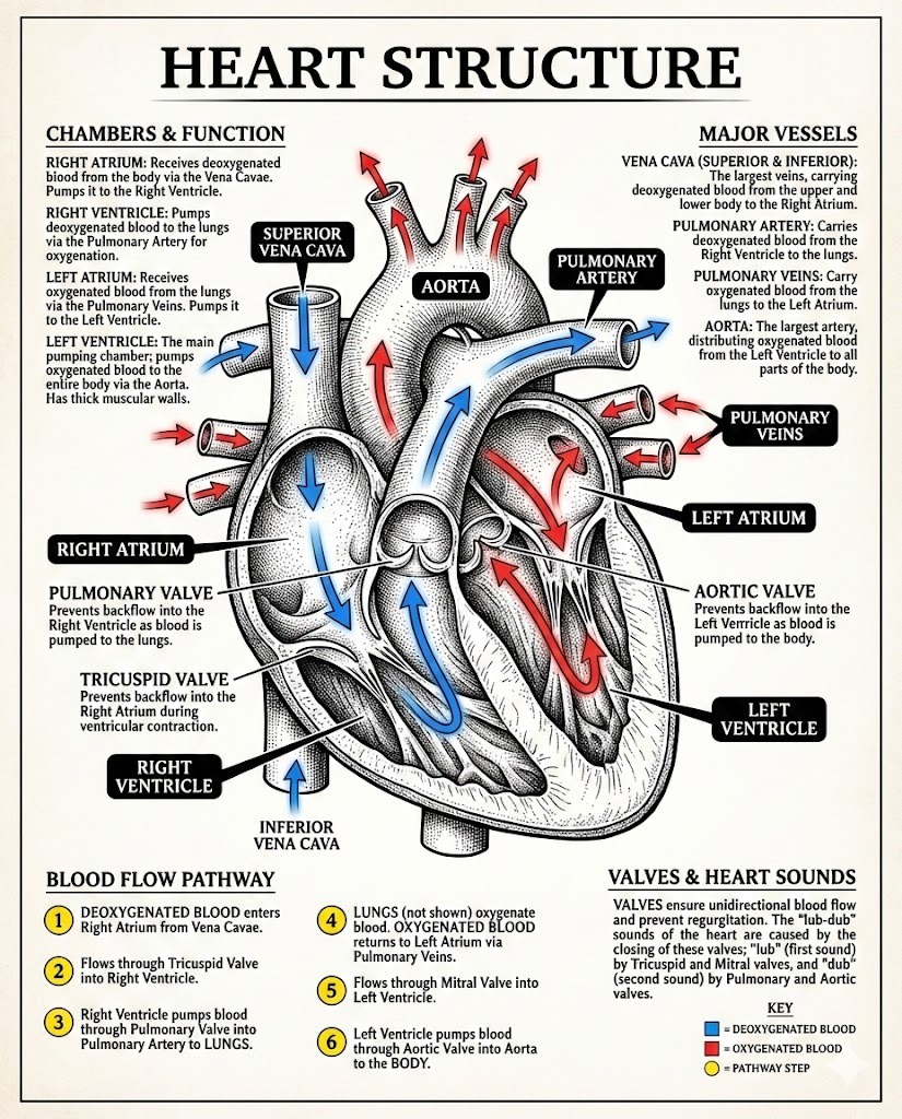
1. Right Atrium
-
Receives deoxygenated blood from the body
-
Blood enters through the superior and inferior vena cava
2. Right Ventricle
-
Pumps deoxygenated blood to the lungs
-
Blood exits via the pulmonary artery
3. Left Atrium
-
Receives oxygenated blood from the lungs
-
Blood enters through pulmonary veins
4. Left Ventricle
-
Pumps oxygenated blood to the entire body
-
Has the thickest muscular wall for powerful pumping
Septa of the Heart
-
Interatrial septum – separates the two atria
-
Interventricular septum – separates the two ventricles
These prevent mixing of oxygenated and deoxygenated blood.
Heart Valves
Valves ensure one-way flow of blood.
Atrioventricular Valves
-
Tricuspid valve – between right atrium and right ventricle
-
Bicuspid (Mitral) valve – between left atrium and left ventricle
Semilunar Valves
-
Pulmonary valve – between right ventricle and pulmonary artery
-
Aortic valve – between left ventricle and aorta
Heart Wall Layers
The heart wall has three layers:
-
Epicardium – outer protective layer
-
Myocardium – thick muscular middle layer (responsible for contraction)
-
Endocardium – inner smooth lining
Blood Supply to the Heart
The heart muscle receives oxygen and nutrients through:
-
Coronary arteries
-
Waste removal via coronary veins
Electrical Conducting System (Brief)
The heartbeat is controlled by a specialized system:
-
SA node (natural pacemaker)
-
AV node
-
Bundle of His
-
Purkinje fibers
This system coordinates rhythmic contractions.
Importance of Heart Structure
The specialized structure of the heart:
-
Prevents blood mixing
-
Maintains efficient circulation
-
Supports continuous pumping without fatigue
Conclusion
The human heart’s structure is perfectly adapted for its vital role in circulation. Its chambers, valves, walls, and vessels work together to ensure uninterrupted blood flow throughout life.