Understanding the structure of animal cells is fundamental to biology, medicine, and biotechnology. Every tissue, organ, and biological system in animals is built from these microscopic units. A clear structural overview of an animal cell helps explain how life functions at its most basic level and how complex organisms maintain growth, repair, and balance.
Animal cells are eukaryotic cells, meaning they contain a nucleus and specialized membrane-bound organelles. Each structure inside the cell performs a specific function that contributes to the survival and operation of the organism. This article provides a comprehensive and accessible explanation of the components that form the animal cell and how they work together as an integrated system.
What Defines an Animal Cell
Animal cells differ from plant cells in several important ways. They lack a rigid cell wall, chloroplasts, and large central vacuoles. Instead, animal cells rely on flexible membranes and specialized internal structures to maintain shape, generate energy, and carry out biological processes.
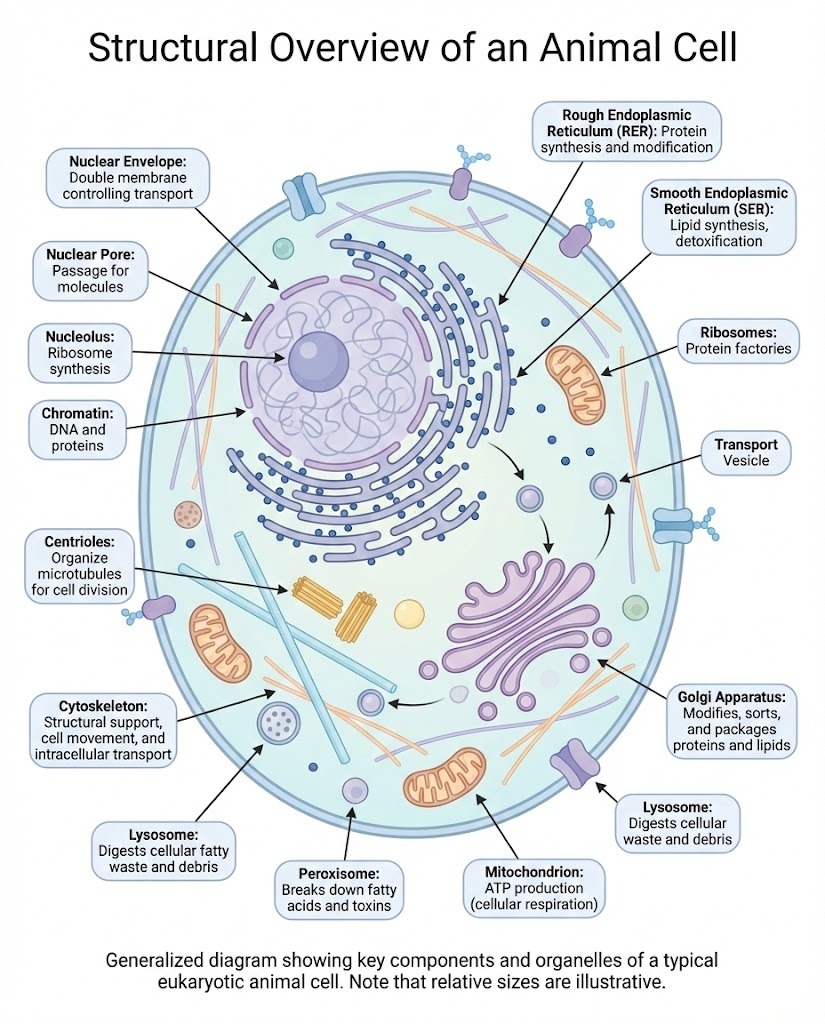
A structural overview of an animal cell typically includes the plasma membrane, nucleus, cytoplasm, and a wide range of organelles suspended within the cytosol. Together, these elements create a highly organized environment where chemical reactions and genetic regulation occur efficiently.
The Plasma Membrane
The plasma membrane forms the outer boundary of the animal cell. It is composed of a phospholipid bilayer embedded with proteins, cholesterol, and carbohydrates.
Key functions of the plasma membrane include:
-
Regulating what enters and exits the cell
-
Maintaining internal balance
-
Enabling cell-to-cell communication
-
Anchoring the cytoskeleton
-
Supporting receptor signaling pathways
Its semi-permeable nature allows essential molecules such as oxygen and glucose to pass while preventing harmful substances from entering.
Cytoplasm and Cytosol
The cytoplasm refers to everything inside the cell membrane except the nucleus. It includes the cytosol, organelles, and cytoskeleton.
The cytosol is a gel-like fluid composed mostly of water, enzymes, ions, and organic molecules. It provides the medium where metabolic reactions take place and allows organelles to remain suspended and properly positioned.
The Nucleus
The nucleus serves as the control center of the cell. It stores DNA and coordinates essential processes such as growth, metabolism, and reproduction.
Major structural components of the nucleus include:
-
Nuclear envelope, a double membrane that protects genetic material
-
Nuclear pores, which regulate transport of RNA and proteins
-
Nucleolus, responsible for ribosome production
-
Chromatin, composed of DNA and proteins
In any structural overview of an animal cell, the nucleus is central because it governs gene expression and cell division.
Mitochondria
Mitochondria are often referred to as the powerhouses of the cell. They generate adenosine triphosphate, the molecule that provides energy for cellular activities.
Important characteristics of mitochondria:
-
Double membrane structure
-
Inner membrane folds called cristae
-
Own circular DNA
-
Ability to replicate independently
Their structure allows efficient energy production through oxidative phosphorylation.
Ribosomes
Ribosomes are responsible for protein synthesis. They can be found floating freely in the cytosol or attached to the rough endoplasmic reticulum.
Free ribosomes generally produce proteins used within the cell, while bound ribosomes create proteins destined for secretion or membrane insertion.
Endoplasmic Reticulum
The endoplasmic reticulum is a network of membranes involved in protein and lipid production.
It consists of two main types:
Rough endoplasmic reticulum
-
Covered with ribosomes
-
Synthesizes proteins
-
Modifies protein structure
-
Prepares proteins for transport
Smooth endoplasmic reticulum
-
Lacks ribosomes
-
Produces lipids and steroids
-
Detoxifies harmful substances
-
Stores calcium ions
Both forms contribute significantly to cellular organization and metabolism.
Golgi Apparatus
The Golgi apparatus acts as the cell’s packaging and distribution center. It receives proteins and lipids from the endoplasmic reticulum, modifies them, sorts them, and directs them to their final destinations.
Functions include:
-
Protein modification
-
Lipid processing
-
Formation of secretory vesicles
-
Production of lysosomes
Its flattened membrane stacks make it easily identifiable in microscopic images.
Lysosomes
Lysosomes contain powerful digestive enzymes that break down waste materials, damaged organelles, and invading microorganisms.
Their roles include:
-
Recycling cellular components
-
Destroying pathogens
-
Supporting programmed cell death
-
Maintaining cellular cleanliness
Without lysosomes, toxic materials would accumulate and damage the cell.
Peroxisomes
Peroxisomes assist in breaking down fatty acids and neutralizing harmful byproducts such as hydrogen peroxide. They play a protective role by reducing oxidative damage within the cell.
Cytoskeleton
The cytoskeleton provides structural support and allows the cell to maintain its shape, transport materials, and divide properly.
It consists of:
-
Microfilaments for movement and tension
-
Intermediate filaments for stability
-
Microtubules for transport and spindle formation
Together, these components form a dynamic internal framework.
Centrosome and Centrioles
The centrosome is the main microtubule-organizing center in animal cells. It contains two centrioles arranged at right angles.
During cell division, centrioles help organize the mitotic spindle that separates chromosomes into daughter cells.
How These Structures Work Together
A structural overview of an animal cell is not complete without understanding coordination. Organelles communicate constantly through chemical signals and transport vesicles. Energy from mitochondria powers protein synthesis in ribosomes, which depends on genetic instructions from the nucleus. Waste products are removed by lysosomes, while membranes are continuously renewed by the endoplasmic reticulum and Golgi apparatus.
This interconnected system allows cells to adapt, respond to environmental changes, and perform specialized functions within tissues.
Importance in Medicine and Research
Understanding animal cell structure is critical for:
-
Diagnosing genetic disorders
-
Developing pharmaceuticals
-
Studying cancer progression
-
Advancing regenerative medicine
-
Designing targeted therapies
Many diseases result from malfunctioning organelles or disrupted cellular organization. A detailed structural understanding enables scientists to pinpoint causes and develop treatments.
Conclusion
A thorough structural overview of an animal cell reveals a remarkably complex and efficient system. Each organelle plays a specialized role, yet none operates in isolation. Together, they support metabolism, growth, communication, and survival.
From the protective plasma membrane to the energy-producing mitochondria and the genetic control center within the nucleus, every component contributes to the greater function of the organism. Appreciating this intricate architecture is essential for anyone studying biology, medicine, or biotechnology.
Frequently Asked Questions About the Structural Overview of an Animal Cell
What is included in a structural overview of an animal cell?
A structural overview of an animal cell includes the plasma membrane, nucleus, cytoplasm, mitochondria, ribosomes, endoplasmic reticulum, Golgi apparatus, lysosomes, cytoskeleton, and centrosome. Each structure has a specific function that supports cellular life.
How does the structure of an animal cell differ from a plant cell?
Animal cells lack a cell wall, chloroplasts, and a large central vacuole. They rely on flexible membranes and different organelle arrangements to perform essential biological processes.
Why is understanding animal cell structure important?
Knowing the structure helps scientists understand how cells function, how diseases develop, and how treatments can target specific cellular components. It is foundational to medical research and biotechnology.
What organelle is most important in the animal cell?
No single organelle is most important. The nucleus, mitochondria, and endoplasmic reticulum are especially critical, but the cell depends on the coordinated function of all its structures to survive and operate effectively.